Probe tips are used in the Atomic Force Microscope (AFM) to detect the surface topography of small objects. A probe is made up of three parts. The substrate is the base of the probe and is the most visible part. The cantilever is the bridge connecting the substrate to the tip. The cantilever controls the tapping frequency of the tip and is barely visible. The tip is the part of the probe that makes contact with the surface and is not visible to the naked eye. A laser shines on the tip, it reflects onto a mirror and then onto a sensor. Finally we get the results of the object’s surface depth, and smoothness.
Probe tips are generally made out of silicon nitride, gold, or platinum. In order to use the probes in the Atomic Force Microscope, the probe must first be inserted into a holder with tweezers. Then the holder is then inserted into the microscope. Probe tips are sensitive and delicate. If a probe tip drops on the floor (or any surface, for that matter) the tip will break. When the tip is broken, it cannot scan the surface of an object. It is almost impossible to insert the probe into the holder without dropping it unless a person has practiced for hours. Dropping a probe automatically means breaking the tip and wasting $50 to $1000.
About five months ago I and my friends Kate Wong, Tiffany Loi, and Winnie Li practiced installing probes in holders. Now that we have to start our own projects, we have to install probes once again. Without practice in five months and without an empty holder to practice with, someone had to take the chance to install the probe. I took the chance, and failed horribly. Afterwards, we had to go apologize to our professor Dr. Nakarmi. When we looked up the cost of the probe I broke … well, let’s just say the number wasn’t pretty.
The SEM images below show three probes with broken cantilevers and tips. We didn’t break all of them. Two of the tips were broken by other people working in the lab.
 |
 |
 |
| One of the broken probes. This side faces the sample being investigated. The probe tip would normally project 0.1 mm out to the left. The numbers inscribed on this one are too small to be visible. | Magnification of the previous image showing the crevice where the cantilever attaches to the probe mount. | The back of the first probe. This side is fixed to the microscope. The sample moves under the the cantilever in two dimensions to create an image of the surface. |
 |
 |
 |
| A broken gold probe. This one does not seem to have a crevice where the cantilever once was. | The back of the second probe. | The last of the broken probes. The edges are chipped away due to practice using tweezers. The chipped edges are not visible in its true size. |
 |
 |
 |
| The back of the third probe showing the alignment grooves. | Close up of the alignment grooves. Because of an optical illusion, the grooves look more like projections. | A mysterious "black hole" on the back of the third probe which is not visible in its true size. |
Image credit: Kate Wong, Tiffany Loi, Yao Jiang, and Winnie Li. Text credit: Yao Jiang (with help from Kate Wong).
Research Coordinator’s supplement …
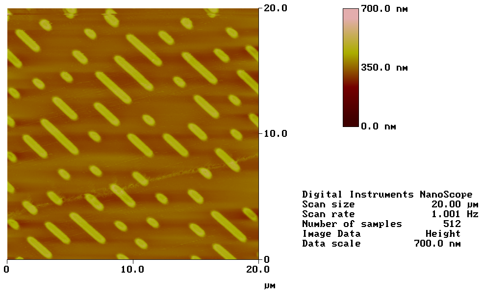
An AFM image of the surface of a DVD made by Ken Han Chen and Chi Vein Cheng in January 2011.
This image was used to make one of the banners for the midwoodscience.org website.