Last week’s SEM images showed the shell of a blue mussel (Mytilus edulis), focusing on the structure of the hard outer shell made of the mineral aragonite (a polymorph of calcium carbonate or CaCO3). This week we’ll be looking at the outside of that same mussel, focusing on a group of microscopic algae whose hard outer shells are made of silicate (also known as hydrated silicon dioxide or H4SiO4).
The little creatures you see below are single celled algae called diatoms. The origin of the word diatom comes from the fact that their shells (called tests or frustules) are made of two interlocking halves. In Greek, dia (διά) means "across" (the diameter of a circle is the measure across it) and tomos (τομος) means "to cut" (atoms are things that can’t be cut), thus diatomos (διάτομος) in Greek or diatoms in English are things that can be "cut across" or "cut in two". Diatoms reproduce asexually by splitting in half. The top half (called the the epitheca) becomes one daughter and the bottom half (called the hypotheca) becomes another. More Greek. The word theca (θήκη) means "case", epi (ἐπί) means "on top", and hypo (ὑπό) means "beneath". Thus, the epitheca (ἐπίθήκη) is the "top case" and the hypotheca (ὑπόθήκη) is the "bottom case".
Diatoms have a light golden brown color due to the presence of chlorophyll a (a green photosynthetic pigment) and chlorophyll c (a yellow photosynthetic pigment). Compare this to trees, grasses, and the other large plants we see around us every day. The leaves of these plants are mostly chlorophyll a and a little bit of chlorophyll b (another yellow photosynthetic pigment). Trees and grasses appear green because the leaves are higher in chlorophyll a than chlorophyll b — 3:1 being a typical a:b ratio. Diatoms appear golden brown because they contain mixtures of chlorophyll a and chlorophyll c that are closer to being equal — a:c ratios from 2:1 (mostly green) all the way to 1:2 (mostly yellow) are found.
Silicon dioxide (SiO2) from sand is the primary raw material for nearly all commercially produced glasses (other ingredients include calcium oxide and sodium carbonate). It is also the primary raw material for the silicate shells of diatoms (the other ingredient is water). In essence, diatoms live in glass houses. Trying to image them with a light microscope is a real challenge. Their clear bodies nearly vanish in the clear liquid they live in. The only thing that makes them stand out is the little bit of yellow-brown pigment in their chloroplasts. To an electron beam, however, these glass housed microalgae are solid as a rock. Light goes through diatoms, but electrons bounce off. A scanning electron microscope is the perfect tool for imaging diatoms and diatoms are the perfect subject for the scanning electron microscope. Enjoy this week’s images and expect to see more diatoms in the future.
 |
 |
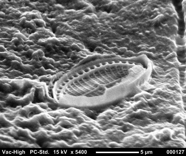 |
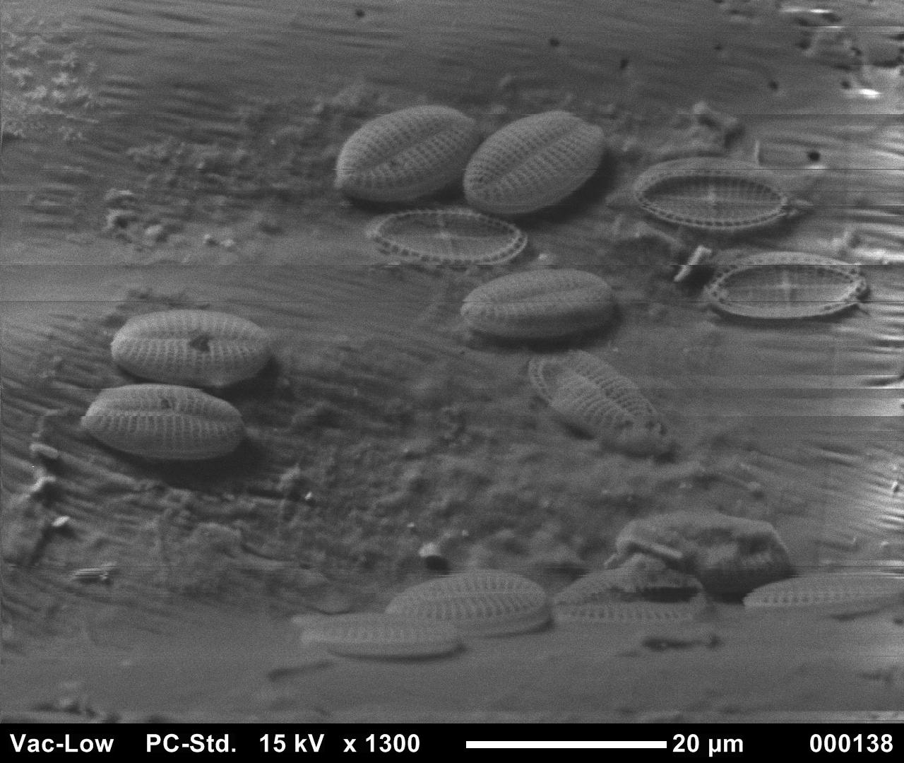
| A group of diatoms hanging out together on the back of a blue mussel. The width of this image is about the same as the width of a human hair. The wavy appearance is an artifact that commonly occurs at high magnification with non-conducting materials. All SEM images are made in a vacuum. | Two whole diatoms with their top half (epitheca) showing and one with its top half missing. These diatoms are in the genus Cocconeis. Possibly Cocconeis scutellum or Cocconeis stauroneiformis. | The bottom half (hypotheca) of one diatom. The top half was blown away by the electron beam. The two halves became negatively charged, like charges repel, and the top half took off. |
Image credit: YaQun Zhou and Anastasiya Matveyenko (images 1 and 2); Glenn Elert (image 3). Thanks to Professors John Marra and Brett Branco at Brooklyn College and Professor Edward Theriot at the University of Texas at Austin for help in identifying these creatures.